RUSTON, La. -- Technology is a fast-moving, fascinating thing. And Louisiana Tech University is using the latest technology to train students in the biological sciences.
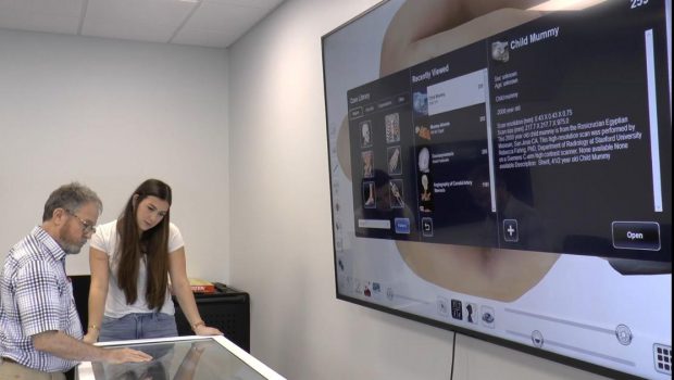
The Justin and Jeanette Hinckley Virtual Anatomy Lab in Carson Taylor Hall now has what is called the Anatomage Table. It is a 3D anatomy and physiology tool that teaches students through hands-on virtual dissection training.
The table allows students to study anatomy and physiology through every system of the body down to the cellular level. It does not use cartoon or computer-generated pictures. These are images of actual people who donated their bodies to science.
“They are real MRI images, real CT scans, real X-ray images,” said William Campbell, the director of the Louisiana Tech School of Biological Sciences. “And so they have been assembled, kind of taken apart from the individual, and then reassembled in the individual. So, you can scroll through these things looking at the different images, the different planes that are represented by the different images. But yes, it's all real data from real people.”
“It is insane, the amount of detail that it can show regarding different systems tissues skin, like everything anatomy wise,” Taylor Teach, a biology graduate student, said about the Anatomage Table. “And I think that overall, it will really enrich Tech's biology curriculum."
The table also contains study material from animals, which will benefit students in pre-vet or animal sciences curricula.
Each body in the table is unique.
“Some of these individuals that are contained in the images that are in the table, these people had different pathologies. And you can see those different pathologies whether it was a tumor or leukemia,” said Campbell. “In the case of some of the animals, there's car accidents, there's gunshot wounds. So, there's a lot to see here, not just the anatomy but pathologies and accidents and that kind of thing.”
Teach says the layers of detail in the table are extraordinary.
“I think the most fascinating part about this table is the scalpel tool, because you can cut into a specific section of the human body and see it, and then tap it to take it apart, layer by layer,” she said.
The Hinkley Virtual Anatomy Lab will be used by multiple departments including biology, nursing, kinesiology, animal sciences and others.


Gloss