Extreme closeup of mouse-brain slice wins top Life Science Microscopy prize
Ainara Pintor/Olympus
For several years now, we've regularly featured the winners of Nikon's annual Small World microscopy contest. Now, Olympus has entered the artful imaging arena with its first Global Image of the Year Award. Like the Small World contest, the intent is to highlight artful scientific imaging in hopes of inspiring the world to appreciate the inherent beauty of microscopy imaging. Olympus announced the winners (one global winner, plus three regional winners), along with several runners-up, last month. They do not disappoint.
As Ars' John Timmer noted in his 2018 Small World coverage: "Microscopy is a sibling of photography in many ways beyond the involvement of high-end lenses. While it might not matter for scientific purposes, a compelling microscope image depends on things like composition, lighting, exposure, and more. And these days, both fields rely heavily on post-processing." All those elements are abundant in the new crop of Olympus winners.
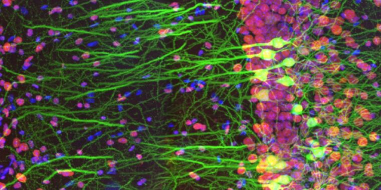
Spain's Ainara Pintor snagged the top honor from over 400 submissions with her gorgeous image of an immunostained mouse-brain slice, titled Neurogarden. The image focuses on the hippocampus area of a single slice, but there are more than 70 million neurons in the mouse brain as a whole, according to Pintor. Howard Vindin of Australia won the regional prize for Asia-Pacific by capturing an autofluorescence image of a mouse embryo. US entrant Tagide de Carvalho won the regional award for the Americas with his colorful image of a tardigrade. The regional winner for Europe, the Middle East, and Africa was the UK's Alan Prescott, for his image capturing the frozen section of a mouse's head.
Honorable mentions included striking microscopic images of photonic crystals in insect scales, crystallized amino acids, desert locust wings, and opal embedded in iron sandstone, among others. Clearly, the field of photomicroscopy is still attracting top-notch talent.
-
Global Winner: immunostaining of a mouse brain slice with two fluorophores
Ainara Pintor/Olympus -
Regional Winner for the Americas: it's a colorful tardigrade!
Tagide de Carvalho/Olympus -
Asia-Pacific Regional Winner: Autofluorescence image of a mouse embryo.
Howard Vindin/Olympus -
EMEA Regional Winner: frozen section of a mouse's head.
Alan Prescott/Olympus -
Honorable Mention: mouse spinal cord.
Tong Zhang/Olympus -
Honorable Mention: image of 3D depth color-coded reconstruction of confocal images of microtubules in monkey fibroblast cells.
Daniela Malide/Olympus -
Photonic crystals in insects (beetles and weevils) FTW!
Rudolf Buechi/Olympus -
Preparation of amino acids crystallized out of an ethanol solution.
Justin Zoll/Olympus -
The desert locust is one example of an insect species that has evolved foldable wings, the better to keep them clean and intact. This image called "A Road in the Sky" because "veins look like roads and spines on [the] wing membrane are like stars."
Hamed Rajabi/Olympus -
Green gem material, prase opal, magnified through a microscope, looks like an aerial coastline view. The brown areas are iron sandstone host rock.
Nathan Renfro/Olympus -
Microscopic image of fruit fly brain
Martin Hailstone/Olympus -
Inflorescence of developing flower buds expressing fluorescent reporters (cyan), with cell walls stained red.
Nat Prunet/Olympus -
The ovary of a gall-inducing wasp showing the eggs.
Ming-Der Lin/Olympus


Gloss